- International medical Journal & Association for Physicians
Синупрет® часто используется в качестве растительного лекарственного средства для лечения синусита, и предполагалось, что противовоспалительное действие может способствовать его общим полезным свойствам. Здесь мы исследовали эффекты лекарственной смеси Sinupret® (SIN), а также нового сухого экстракта Синупрета (SIN DE) с последним, содержащим более высокие концентрации активных ингредиентов, в модели острого воспаления in vivo, каррагинан-индуцированный плеврит у крыс и SIN, и SIN DE вводили крысам перорально в дозах 100 мг / кг (низкая доза) и 500 мг / кг (высокая доза) за 1 ч. до внутриплевральной инъекции каррагинана. Хотя как SIN, так и SIN DE значительно снижали объем экссудата и количество лейкоцитов в плевральном экссудате при высокой и низкой дозе через 4 часа после инъекции каррагинана, новый SIN DE был более эффективен, чем SIN при низкой дозе, что подразумевало более высокую эффективность. Параллельно с этим новый сухой экстракт SIN DE, но не SIN, в дозе 500 мг/кг значительно снижал уровни простагландина (PG) E2 в экссудатах и уменьшал количество белка циклооксигеназы (COX) -2 в легких. Вместе SIN и SIN DE оказывают существенное противовоспалительное действие при пероральном приеме, что позволяет рационально использовать их для лечения синусита и других вирусных / микробных назальных инфекций, связанных с воспалением. Более того, наши результаты показывают, что, основываясь на более высокой эффективности и сопровождающемся снижении экспрессии COX-2 и образовании PG E2, новый сухой экстракт SIN DE может быть лучше, чем прежняя лекарственная смесь SIN.
The novel Sinupret® dry extract exhibits anti-inflammatory effectiveness in vivo
ABSTRACT
Sinupret® is frequently used as a herbal medicinal product to treat sinusitis, and it was assumed that anti-inflammatoiy effects might contribute to its overall beneficial properties. Here, we investigated the effects of a Sinupret® drug mixture (SIN) as well as of the novel Sinupret® dry extract (SIN DE) with the latter containing higher concentrations of active ingredients, in an in vivo model of acute inflammation, the carrageenan-induced pleurisy in rats. Both SIN and SIN DE were administered to rats orally at doses of 100 mg/kg (low dose) and 500 mg/kg (high dose) 1 h prior to intrapleural injection of carrageenan. Although both SIN and SIN DE significantly reduced the exudate volume and leukocyte numbers in the pleural exudate at the high and the low dose 4 h after carrageenan injection, the novel SIN DE was more efficient than SIN at the low dose, implying higher efficiency. In parallel, the novel dry extract SIN DE, but not SIN, at 500 mg/kg significantly lowered the levels of prostaglandin (PG) E2 in the exudates and reduced the amounts of cyclooxygenase (COX)-2 protein in the lungs. Together, SIN and SIN DE exert significant oral anti-inflammatory effects, which rationalize their therapeutic use in the management of sinusitis and other viral/microbial nasal infections that are associated with inflammation. Moreover, our results suggest that based on the higher efficiency and the accompanied reduction of COX-2 expression and PCE2 formation, the novel dry extract SIN DE might be superior over the former SIN drug mixture.
1. Introduction
Inflammatory diseases are accompanied by neutrophil in-filtration, cytokine release (e.g., tumor necrosis factor (TNF)a and interleukin (IL)-lp) and formation of pro-inflammatory mediators (e.g., leukotrienes (LT) and prostaglandins (PGs)). Several experimental Findings have shown that these mediators contribute to tissue damage characteristic of the inflammatory process in vivo [1-5]. In addition, activated neutrophils release various proteases (e.g., leukocyte elastase or cathepsin G) and generate reactive oxygen species [6-8], both of which destroy invading particles but also damage cells and tissues of the host. Interference with the generation or action of these pro-inflammatory mediators exerts beneficial effects in a variety of inflammation models including the carrageenan-induced pleurisy model [9,10].
Sinupret® is a herbal medicinal product composed of Gentianae radix, Primulae flos cum calycibus, Sambuci flos, Rumicis herba (sorrel), and Verbenae herba that is frequently used for the treatment of acute or chronic rhinosinusitis in Germany and other European countries [11 ]. In carrageenan- induced paw edema studies an anti-inflammatory effect of Sinupret® as well as one of its herbal ingredients could be shown [12,13]. Moreover, patients with acute otitis media daily treated three times with two tablets of Sinupret® for ten days improved the disease state accompanied by a slight drop of the pro-inflammatory cytokines TNFa and IL-6 levels in the blood [14]. Based on a highly standardized dry extract of the well-established combination of five herbal drugs, a novel Sinupret® herbal medicinal product was developed that contains higher concentrations of active ingredients.
Here we investigated the ability of the Sinupret® drug mixture (SIN) and the newly developed Sinupret® dry extract (SIN DE) to suppress typical pro-inflammatory events in a well- established and robust in vivo model of acute inflammation, that is, the carrageenan-induced rat pleurisy, which is widely utilized for the in vivo screening of anti-inflammatory compounds [9]. Our data show that both, SIN and SIN DE are efficient anti-inflammatory agents, while the novel SIN DE has superior efficiency over the drug mixture, and it also significantly inhibits PGE2 synthesis and cyclooxygenase (COX)-2 expression. We conclude that the newly developed dry extract SIN DE possesses a promising pharmacological profile supporting its potential therapeutic application.
2. Experimental
2.1. Materials
SIN (BNO D_100) was supplied by the manufacturer. The mixture contains pulverized drugs from Gentianae radix, Primulae flos cum calycibus, Sambuci flos, Rumicis herba (sorrel), and Verbenae herba in the fixed ratio of 1:3:3:3:3. SIN DE, a native dry extract (special extract BNO 1011), was also supplied by the manufacturer. The composition of the native dry extract SIN DE regarding the herbs is identical to that of SIN, however, the dry extract SIN DE contains higher concentrations of active ingredients compared with SIN. SIN DE was prepared with 59% ethanol as extracting agent (V/V) (with a final drug: extract ratio of 4.2:1). The quality of herbal drugs (starting material) is specified according to the relevant EMA-Guidelines for herbal medicinal products. Both, SIN and SIN DE, were manufactured in a validated production process according to GMP. Comprehensive specifications and standardized production processes guarantee high batch-to-batch consistency.
For the analysis of the SIN and of SIN DE in the pleurisy model, the compounds were freshly (not more than 4h prior application) resuspended in water at room temperature using a magnetic stirrer. Homogenous suspensions were obtained and used for further analysis. X-Carrageenan type IV isolated from Cigartina aciculaire and Gigartina pistillata was purchased from Sigma-Aldrich (Milan, Italy). 3H-PGE2 was from PerkinElmer Life Sciences (Milan, Italy) and PGE2 antibody from Sigma-Aldrich (Milan, Italy). Other reagents and compounds used were obtained from Sigma-Aldrich (Milan, Italy) if not stated otherwise.
2.2. Animals
Male Wistar Han rats (220-230 g, San Pietro al Natisone, Italy) were housed in a controlled environment and standard rodent chow and water were provided ad libitum. Animal care complied with Italian regulations on protection of animals used for experimental and other scientific purpose (Ministerial Decree 116192) as well as with the European Economic Community regulations (Official Journal ofE.C. L 358/1 12/18/ 1986).
2.3. Carrageenan-induced pleurisy
Rats were divided into six groups (n = 10 for each group). SIN and the novel SIN DE (100 and 500 mg/kg, each), indo- methacin (5 mg/kg) or vehicle (1 mL water) were given per os 1 h before carrageenan. Rats were anaesthetized with enflurane (4%) mixed with 02 (0.5 L/min), N20 (0.5 L/min), and submitted to a skin incision at the level of the left sixth intercostal space. The underlying muscle was dissected, and saline (0.2 mL) or 1% -carrageenan type IV (w/v) (0.2 mL) was injected into the pleural cavity. The skin incision was closed with a suture, and the animals were allowed to recover. 4 h after the injection of carrageenan, the animals were killed by inhalation of C02. The chest was carefully opened, and the pleural cavity was rinsed with 2 mL saline solution containing heparin (5U/mL). The exudates and washing solution were removed by aspiration, and the total volume was measured. Any exudate that was contaminated with blood was discarded. Polymorphonuclear neutrophils (PMNs) in the exudates were resuspended in phosphate-buffered saline (PBS) and counted with an optical light microscope in a Burker's chamber after vital trypan blue staining. The exudates were centrifuged at 800 xg for 10 min. The amount of LTB4 and PGE2 in the supernatant was assayed by enzyme immunoassay and radioimmu-noassay (Cayman Chemical, Ann Arbor, MI), respectively, according to manufacturer's protocol. TNFa and IL-ip levels were evaluated in the supernatants by using ELISA kits (R & D Systems, Minneapolis, USA). The results are expressed as nanogram per rat.
2.4. Analysis of COX-2 expression in rat lungs
At 4 h after carrageenan administration, the lungs were ho-mogenized in a buffer containing 20 mM HEPES pH 7.6,1.5 mM MgCl2, 400 mM NaCl, 1 mM EDTA, 1 mM EGTA, 1 mM dithio- threitol, 0.5 mM phenylmethylsulfonyl fluoride, 15|jg/mL trypsin inhibitor, 3 (Jg/ml pepstatin, 2 pg/mL leupeptin, 40 |JM benzamidine, 1% Nonidet P-40, 20% glycerol, and 50 mM NaF. The homogenates were centrifuged at 10,000 xg for 15 min at 4 °C, the supernatant was collected, and protein concentration was determined by Bio-Rad protein assay (Bio-Rad Laboratories, Milan, Italy). Equal amounts of protein (30 (Jg) were mixed with gel loading buffer (50 mM Tris, 10% SDS, 10% glycerol, 10% 2-mercaptoethanol, and 2 mg/mL bromophenol) in a ratio of 1:1, boiled for 3 min and centrifuged at 10,000 xg for 10 min. Each sample was loaded and electrophoresed on a 10% SDS-polyacrylamide gel. The proteins were transferred onto nitrocellulose membranes. The membranes were blocked with 0.1% PBS-Tween containing 5% non-fat dry milk. After the blocking, the membranes were incubated with the primary antibody for 2 h at room temperature. Rabbit monoclonal antibody anti-COX-2 (Cayman Chemical, Ann Arbor, Ml) was diluted 1:500 in 0.1% PBS-Tween containing 5% non-fat dry milk. After the incubation, the membranes were washed six times with
0.1% PBS-Tween and were incubated for 1 h at room temperature with horseradish peroxidase-conjugated secondary anti-body (Dako Cytomation, Glostrup, Denmark) diluted 1:1000 in 0.1% PBS-Tween containing 5% non-fat dry milk. The membranes were washed and protein bands were detected by an enhanced chemiluminescence system (GE Healthcare, Freiburg, Germany). Densitometric analysis was performed with a Fluor S quantitative imaging system (Bio-Rad Laboratories, Milan, Italy).
2.5. Statistical procedures
All values in the figures and text are expressed as mean ± standard error of the mean (SEM) of n observations, where n represents the number of animals studied. The data were ana-lyzed by one-way ANOVA followed by Tukey HSD post-hoc tests. A P value less than 0.05 was considered statistically significant.
3. Results
3.1. Inhibition of exudate formation and PMN infiltration in carrageenan-induced pleurisy
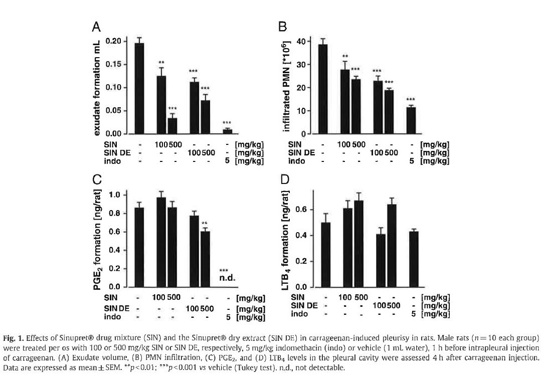
To assess the effectiveness of SIN and of SIN DE as anti-inflammatory agents in vivo, the compounds were tested in a severe model of acute inflammation, that is, pleurisy induced by carrageenan in rats. Injection of carrageenan into the pleural cavity of rats (vehicle group, 10 animals per group) elicited an acute inflammatory response within 4 h characterized by the accumulation of fluid that contained large numbers of PMNs (Fig. 1A/B). Per os pre-treament of rats (1 h) with 100 or 500 mg/kg SIN or SIN DE inhibited the inflammatory response induced by intrapleural injection of carrageenan, as demonstrated by the significant attenuation of exudate formation and PMN infiltration, respectively (Fig. 1A/B). Note that SIN DE was more efficient than SIN at the low dose regarding the reduction of both exudate formation and PMN infiltration. As expected, the potent non-steroidal anti-inflammatory drug (NSAID) indomethacin (5 mg/kg, per os), used as reference drug, strongly suppressed the inflammatory response as well (Fig. 1A/B).
3.2. Analysis of eicosanoid levels in pleural exudates of carrageenan-treated rats
The eicosanoids, i.e., PG and LT produced by the 5- lipoxygenase and the COX pathway, respectively, are major low molecular weight compounds that mediate numerous pro-inflammatory events [15], and LTB4 and PGE2 were shown to be elevated in the exudates of carrageenan-treated rats [1,16]. SIN failed to affect PGE2 and LTB4 formation in carrageenan-treated rats (Fig. 1C/D). When rats were pretreated with 100 mg/kg SIN DE, the levels of PGE2 were slightly (10%) but not significantly lower compared to vehicle-treated animals. However, at the higher dose of 500 mg/kg, SIN DE led to a significant inhibition ofPGE2 formation (30%; p<0.01;
Fig. 1C). In contrast, the LTB4 levels were not altered by SIN DE (Fig. ID). As expected, the COX inhibitor indomethacin blocked PGE2 synthesis but LTB4 was not affected.
3.3. Analysis of COX-2 expression in the lungs of carrageenan-treated rats
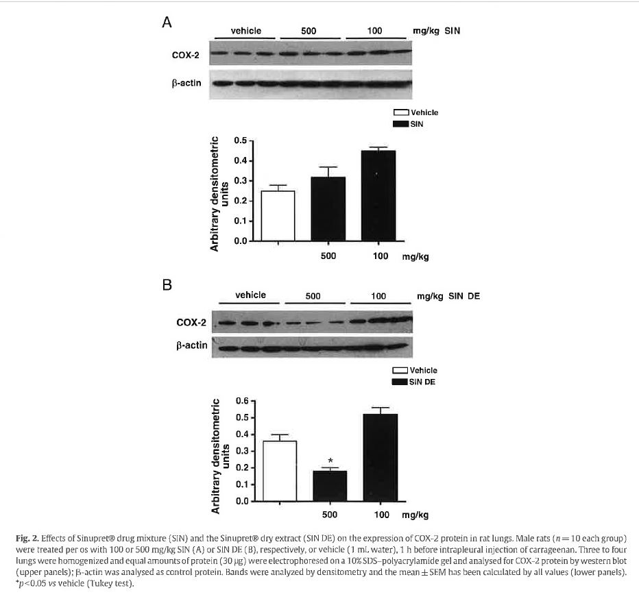
The impaired PGE2 formation in rats treated with high-dose SIN DE encouraged us to investigate whether the extract may affect the protein expression of the key enzyme in PGE2 formation at inflammatory sites, namely COX-2 [15,17]. Western blot analysis of lungs of rats after carrageenan-treatment revealed strong COX-2 protein induction in vehicle-treated animals (Fig. 2). However, in the lungs of rats that received the high dose of SIN DE (500 mg/kg), the COX-2 protein levels were significantly reduced by about 42% (Fig. 2B). For comparison, also lungs of rats pre-treated with SIN were analyzed for COX-2 protein but, in contrast to SIN DE-treated rats, no impaired protein levels were observed (Fig. 2A).
3.4. Analysis of cytokine levels in the exudates of carrageenan- treated rats
The release of inflammatory cytokines, such as TNFa and IL-ip by activated PMN, represents a pivotal biochemical event to initialize, maintain, and propagate the inflammatory response within injured tissues [3]. Pre-treatment of rats with 100 mg/kg SIN or SIN DE impaired the 1L-1(5 levels by 27 and 11% (p>0.05), respectively. At the higher dose (500 mg/kg), IL-1[J was reduced by 40% (p<0.01) and 19%, respectively (Fig. ЗА). In contrast, the carrageenan-induced elevation of TNFa was not affected by either SIN or SIN DE (Fig. 3B). Note that indomethacin caused neither an impairment of TNFa nor of IL-lp, as expected.
4. Discussion
Here, we have assessed the efficacy of SIN drug mixture and of the novel advanced dry extract SIN DE after per os adminis¬tration in a well-established, robust in vivo animal model of acute inflammation, the carrageenan-induced rat pleurisy. Both SIN and SIN DE clearly reduced the inflammatory response, with the dry extract SIN DE being superior over the drug mixture SIN at the low dose of 100 mg/kg. Moreover, our results provide some insights into the biochemical mecha¬nisms. Thus, reduced COX-2 expression and accompanied decreased generation of pro-inflammatory PGE2 may account for the efficient anti-inflammatory effect of SIN DE, whereas impaired IL-ip release seems to contribute to the antiinflammatory action of SIN drug mixture. We conclude that SIN and SIN DE possess anti-inflammatory activity when given orally, where the dry extract SIN DE might be superior over SIN drug mixture, and these properties might contribute to the well-recognized beneficial features in the treatment of sinusitis.
Based on its secretolytic, anti-viral, and anti-bacterial prop-erties, the Sinupret® medicinal product is commonly used as herbal medicine to treat rhinosinusitis. Sinupret® was shown to release tough mucus, to clear the congested nose, to alleviate head pressure and headache, as well as to fight against both viral and bacterial infections [11,12,18]. Moreover, anti-inflammatory features have been proposed to contribute to the overall beneficial properties of the Sinupret® herbal medicinal product [12-14]. Our present data unambiguously confirm such anti-inflammatory properties of the Sinupret® drug mixture SIN in vivo as exudate formation in the pleural cavity and the infiltration of pro-inflammatory PMN after carrageenan- challenge of the rats were significantly and dose-dependently reduced after per os treatment of the rats. Hence, for the first time, we likewise substantiate anti-inflammatory activity in vivo for the novel dry extract SIN DE.
α formation our data also allow to speculate about the mechanisms underlying the anti-inflammatory action of the extracts. The carrageenan-induced pleurisy is accompanied by eicosanoid generation and cytokine release which are considered to be required for induction of the inflammatory response [1-5]. Upon carrageenan challenge, the expression of the inducible COX-2 was strongly upregulated within 4 h and this was accompanied by a strong increase in the formation of PGE2. Among the PGs, PGE2 plays the major role in mediating the inflammatory response, and the anti-inflammatory effectiveness of NSAIDs correlates with reduced PGE2 generation [19]. In fact, the reference compound indomethacin, a well- recognized NSAID that acts by inhibiting COX-1/2 [20], sup-pressed PGE2 down to non-detectable levels, accompanied by almost complete prevention of exudate formation and PMN in-filtration. Note that neither LTB4 levels nor TNFct and IL-lfi release were significantly suppressed by indomethacin, sup-porting the critical role of prostanoids, seemingly produced by COX-2 under the experimental conditions used [17]. High- dose SIN DE blocked COX-2 expression and at the same time significantly impaired the PGE2 levels. Of interest, as found for indomethacin, the levels of LTB4 and TNFa were not reduced by the dry extract SIN DE, and only a minor decrease of IL-1(3 was evident, implying that suppression of PGE2 might essentially underlie the anti-inflammatory effect of SIN DE. On the other hand, also SIN significantly reduced the exudate formation and PMN infiltration but failed to lower COX-2 expression and PGE2 levels. It is possible that some bioactive components) are enriched in SIN DE vs SIN and that they reach sufficient bioactive concentrations only in the high-dose SIN DE group. These enriched bioactive substances may be responsible for the repression of COX-2 and PGE2. Nevertheless, SIN significantly impaired IL-1(J levels, which may be an alternative cause for the anti-inflammatory activity. In fact, in acute (4 h) zymosan-induced rat pleurisy, polyclonal murine anti-IL-1 (3. serum selectively inhibited the number of neutrophils that migrated to the inflamed site [21], and also pre-treatment of rats with a human IL-1JJ receptor antagonist suppressed the infiltration of cells into the pleural cavity during acute (5h) carrageenan-induced pleurisy [22]. At the moment, we can only speculate about the biochemical mechanisms that cause IL-l(i reduction by SIN and COX-2 repression by SIN DE. On one hand, the expression of IL-1(J might be affected by SIN as is the case for glucocorticoids that block IL-1(3> gene transcription involving the nuclear factor-kappaB (NFKB) pathway [23,24]. On the other hand, SIN might interfere with post- translational mechanisms and secretion of IL-lfi. Suppression of COX-2 protein and thus reduced PGE2 synthesis by SIN DE may also be due to several possible mechanisms such as interference with the signalling pathway induced by carrageenan (including the NFKB pathway) [25] or degradation of COX-2 mRNA. Finally, we would like to emphasize that interference with additional pro-inflammatory key events and key molecules (that have not been subject of investigation in this study) may contribute to the anti-inflammatory actions of the extracts, including suppression of other cytokines (e.g., 1L-2, -6, -8, -12, -23), protease secretion, reactive oxygen species formation, inducible nitric oxide (NO) synthase expression, and NO generation, as well as inhibition of histamine release.
In conclusion, both SIN and the dry extract SIN DE exhibited potent anti-inflammatoiy activity during carrageenan-induced pleurisy in vivo after per os treatment of rats. These data support the therapeutic use of Sinupret® against sinusitis, a pathological process that has a strong inflammatory component, and rationalize further in vitro analysis of the biochemical and molecular mechanistic details underlying the anti-inflammatory effects of SIN and SIN DE.
Antonietta Rossia, Friederike Dehm b, Christoph Kiesselbachc, Jutta Haunschildc,
Lidia Sautebina, Oliver Werz b
Journal Fitoterapia 83 (2012), p 715-720
aDepartment of Experimental Pharmacology, University of Naples Federico II, Via D. Montesano 49, 80131 Naples, Italy
bChair of Pharmaceutical/Medicinal Chemistry, Institute of Pharmacy, University of Jena, Philosophenweg 14, D-07743 Jena, Germany
c Bionorica SE, Kerschensteinerstr. 11-15, D-92318 Neumurkl, Germany
References
[1 ] Cuzzocrea S, Rossi A, Serraino I, Mazzon E, Di Paola R, Dugo L, et al. 5- Lipoxygenase knockout mice exhibit a resistance to pleurisy and lung injury caused by carrageenan. J Leukoc Biol 2003;73:739-46.
[2] Fialkow L, Wang Y, Downey GP. Reactive oxygen and nitrogen species as signaling molecules regulating neutrophil function. Free Radic Biol Med 2007;42:153-64.
[3] Frode IS, Souza GE, Calixto JB. The modulatory role played by TNF-alpha and IL-1 beta in the inflammatory responses induced by carrageenan in the mouse model of pleurisy. Cytokine 2001;13:162-8.
[4] Mazzon E, Cuzzocrea S. Role of TNF-alpha in lung tight junction alteration in mouse model of acute lung inflammation. Respir Res 2007;8:75.
[5] Ueno A, Oh-ishi S, Critical roles for bradykinin and prostanoids in acute inflammatory reactions: a search using experimental animal models. Curr Drug Targets Inflamm Allergy 2002;1:363-76.
[6] Conner EM, Grisham MB. Inflammation, free radicals, and antioxidants. Nutrition 1996;12:274-7.
[7] Krause KH, Campbell KP, Welsh MJ, Lew DP. The calcium signal and neutrophil activation. Clin Biochem 1990;23:159-66.
[8] Parekh AB, Penner R. Store depletion and calcium influx. Physiol Rev 1997;77:901-30.
[9] Moore AR. Pleural models of inflammation: immune and nonimmune. Methods Mol Biol 2003;225:123-8.
[10] Morris CJ. Carrageenan-induced paw edema in the rat and mouse. Methods Mo! Biol 2003;225:115-21.
[11] Melzer J, Sailer R, Schapowal A, Brignoli R. Systematic review of clinical data with BNO-lOl (Sinupret) in the treatment of sinusitis. Forsch Komplementmed 2006;13:78-87.
[12] Marz RW, Ismail C, Popp MA. Profile and effectiveness of a phytogenic combination preparation for treatment of sinusitis. Wien Med Wochenschr 1999;149:202-8.
[13] SchwartnerC, Michel C.StettmaierK,WagnerH, BorsW.Marchantinsand related polyphenols from liverwort: physico-chemical studies of their radical-scavenging properties. Free Radic Biol Med 1996;20:237-44.
[14] Chkhaidze I, Nemsadze K, Gotsadze K, Nikoleishvili E, Gordeladze G. Sys¬temic inflammatory responses in patients with acute otitis media and the impact of treatment with Sinupret. Georgian Med News 2007:40-4.
[15] Funk CD. Prostaglandins and leukotrienes: advances in eicosanoid biol¬ogy. Science 2001;294:1871-5.
[16] Rossi A, Di Paola R, Mazzon E, Genovese T, Caminiti R, Bramanti P, et al. Myrtucommulone from Myrtus communis exhibits potent anti- inflammatoiy effectiveness in vivo. J Pharmacol Exp Ther 2009;329: 76-86.
[17] Masferrer JL, Zweifel BS, Manning PT, Hauser SD, Leahy KM, Smith WG, et al. Selective inhibition of inducible cyclooxygenase 2 in vivo is antiin¬flammatory and nonulcerogenic. Proc Natl Acad Sci U S A 1994;91: 3228-32.
[18] Ismail C. Pharmacology of Sinupret. Recent results on the rational for the Sinupret compound. HNO 2005;53{Suppl l):S38-42.
[19] Rainsford KD. Anti-inflammatory drugs in the 21st century. Subcell Bio¬chem 2007;42:3-27.
[20] Ferreira SH, Moncada S, Vane JR. Indomethacin and aspirin abolish prostaglandin release from the spleen. Nat New Biol 1971;231:237-9.
[21] Perretti M, Soli to E, Parente L. Evidence that endogenous interleukin-1 is involved in leukocyte migration in acute experimental inflammation in rats and mice. Agents Actions 1992;35:71-8.
[22] Meyers KP, Czachowski CL, Coffey JW. Effect of treatment with interleukin-1 receptor antagonist on the development of carrageenan- induced pleurisy in the rat. Inflammation 1993;17:121-34.
[23] Knudsen PJ, Dinarello CA, Strom ТВ. Glucocorticoids inhibit transcrip¬tional and post-transcriptional expression of interleukin 1 in U937 cells. J Immunol 1987;139:4129-34,
[24] Ray A, Prefontaine KE. Physical association and functional antagonism between the p65 subunit of transcription factor NF-kappa В and the glucocorticoid receptor. Proc Natl Acad Sci U S A 1994;91:752-6.
[25] D’Acquisto F, Ianaro A, lalenti A, IuvoneT, Colantuoni V, Carnuccio R. Acti¬vation of nuclear transcription factor kappaB in rat carrageenan-induced pleurisy. EurJ Pharmacol 1999;369:233-6.
Elsevier GmbH Professional Solutions
Hackerbrucke 6 • D-80335 Miinchen Tel: +49-(0) 89-5383-704 Fax: +49-(0) 89-5383-725 e-mail: sonderdrucke@elsevier.de www.elsevier.de/professional-solutions
No lesponsibility is assumed by Elsevier, its licensors or associates (or any injury and/or damage lo persons or property as a matter of products liability, negligence or otherwise, or from any use or operation of any methods, products, instructions, or ideas contained in the material herein. Because of rapid advances in the medical sciences, in particular, independent verification of diagnoses and drug dosages should be made.
This article reprint is distributed with the support of:
Bionorica St.
29.01.2020 7426